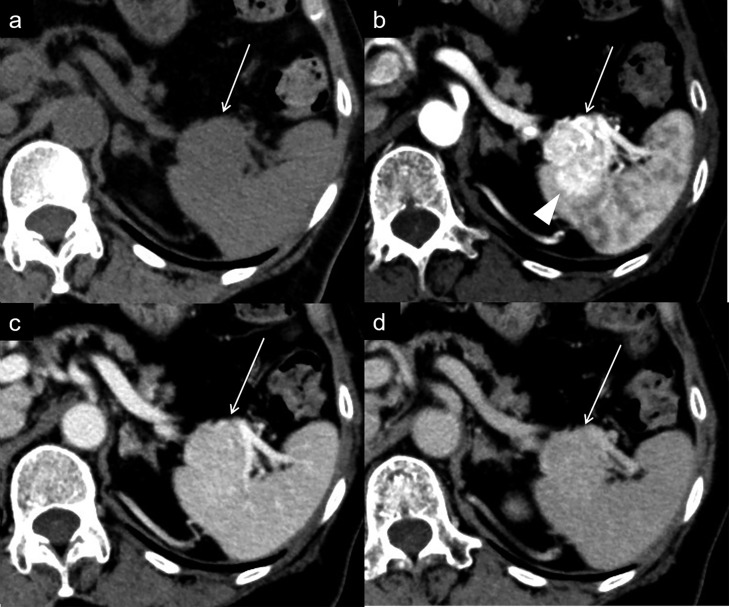
Figure 1.
Dynamic contrast-enhanced CT (a. unenhanced phase, b. arterial phase, c. portal venous phase, d. equilibrium phase). A homogenous mass (a: arrow) is present in the tail of the pancreas. The mass shows heterogeneously marked enhancement in the arterial phase (b: arrow) and washout in the portal venous and equilibrium phases (c, d: arrow). The splenic invasion is clearly identified in the arterial phase (b: arrowhead).

