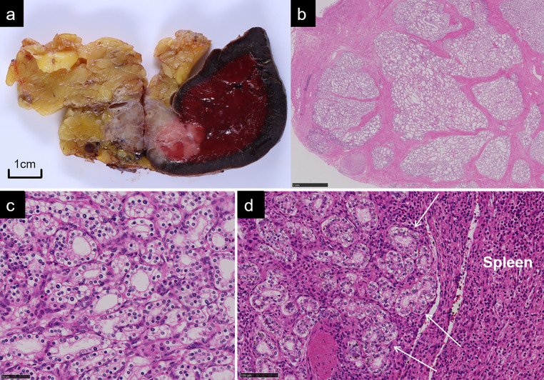
Figure 3.
(a) Formalin-fixed specimen demonstrates a well-circumscribed tumour in the tail of the pancreas with an infiltrative growth into the spleen. Cysts are barely identifiable in the tumour. (b, c, d) Histopathological photomicrographs with hematoxylin and eosin staining shows microcystic appearance lined by epithelial cells with clear cytoplasm (b. low power field, c. high power field) and direct invasion into the splenic parenchyma (d. low power field).

