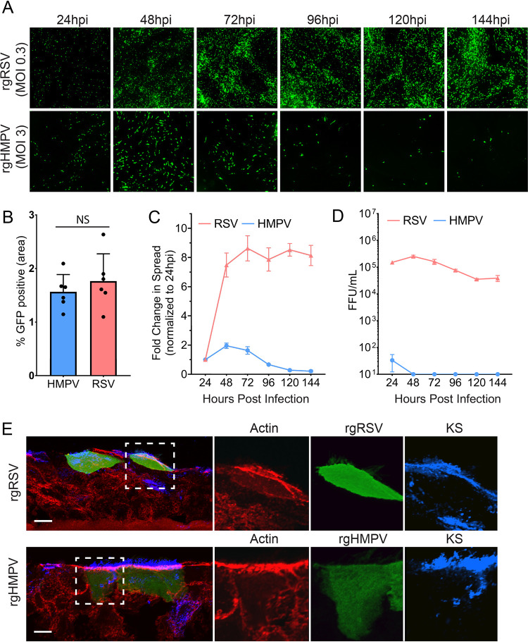
FIG 1.
RSV and HMPV infection, spread, and release in HAE tissues. (A) HAE tissues were infected with MOI 0.3 of rgRSV or MOI 3.0 of rgHMPV, and initial infection and spread were examined up to 144 hours postinfection (HPI). (B) RSV and HMPV infection at the 24 hpi time point. (C) Spread analysis of HMPV and RSV were determined using fluorescence threshold analysis. (D) Apical release of virus was determined by washing the apical surface of HAE tissues, determining the titer of the viral wash in 2-D monolayers, and calculating fluorescence-forming units (FFU). (E) Infected cells for HMPV (48 hpi) and RSV (72 hpi) were stained for actin cytoskeleton, as well as keratan sulfate (KS) to stain for ciliated cells. Error bars represent the standard error of the mean (SEM) of 6 different tissues. Scale bar = 10 μm.

