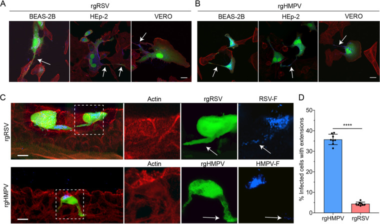
FIG 4.
HMPV forms intercellular extensions significantly more than RSV. (A and B) BEAS-2B, Vero, and HEp-2 cells infected with rgRSV (A) or rgHMPV (B) demonstrate the formation of long filamentous extensions. (C) HAE tissues infected with either rgRSV or rgHMPV demonstrate the formation of these filamentous extensions in a 3-D model system. (D) Extension formation is significantly more common in rgHMPV-infected tissues than in those infected with rgRSV. Statistical significance is represented with P < 0.05 (*), P < 0.005 (**), P < 0.0005 (***), and P < 0.0001 (****). Scale bar = 25 μm for 2-D cell culture, 10 μm for HAE tissues, and 5 μm for higher magnification insets.

