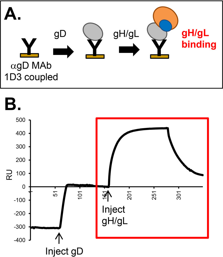
FIG 8.
Demonstration of gH2/gL2 binding to gD2, as detected by SPR. (A) Schematic diagram of the SPR protocol. Anti-gD MAb 1D3, which is permissive for gH/gL binding (21), was coupled to a CM5 biosensor chip. Sequential injections of soluble gD2(285t) and soluble gH2/gL2 were performed. (B) An increase in resonance units (RU) after the gH/gL injection indicated gH/gL binding (red box). The RU from a control flow cell where only gH/gL was injected (no gD) was subtracted.

