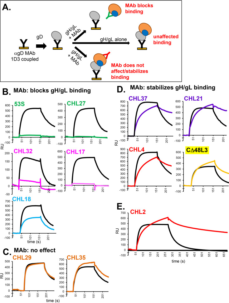
FIG 9.
Effect of anti-gH/gL MAbs on gD-gH/gL binding. (A) Schematic diagram of the SPR protocol. Anti-gD MAb 1D3 was coupled to a CM5 biosensor chip and then soluble gD2(285t) was injected across the flow cell. Next, either gH2/gL2 alone or gH2/gL2 that was preincubated with 0.4 mg/ml of the indicated MAb (IgG) was injected across the flow cell. The RU from a control flow cell where only gH/gL or gH/gL + IgG was injected (no gD) was subtracted from each. (B) MAbs that block gH/gL binding to gD. (C) MAbs that do not affect gD-gH/gL binding. (D) MAbs that stabilize the gD-gH/gL binding. Only the gH/gL binding portion of the curves (Fig. 8B, red box) are shown in parts (B) to (E). Black curves show gH/gL binding to gD when no MAb is added (positive control). Curves where gH/gL was preincubated with MAbs are colored according to their MAb community in Fig. 2B. Each experiment was done at least twice and a representative experiment is shown.

