FIG 4.
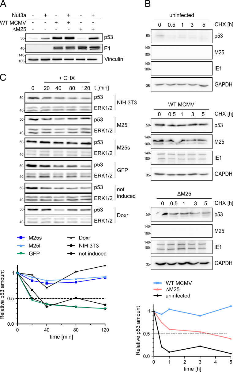
The half-life of p53 is increased in WT MCMV-infected cells and in M25-expressing cells. (A) NIH 3T3 cells were used either uninfected or were infected with WT MCMV or ΔM25 MCMV. At 18 h p.i., the cells were cultivated for another 6 h or treated with Nutlin3a (40 μM), followed by immunoblot analysis of cell lysates with the indicated antibodies. Vinculin served as a loading control, and the viral E1 early protein served as an infection marker. (B) NIH 3T3 cells either uninfected or infected as described previously (see panel A) for 24 h were then treated with cycloheximide (CHX; t = 0 h). At the specified time points thereafter, lysates of cells were prepared and analyzed by immunoblotting with the indicated antibodies. GAPDH served as a loading control, and the viral immediate early protein IE1 was used as an infection marker. (C) Parental NIH 3T3 cells and cell lines encoding the indicated proteins were treated with doxycycline for 24 h, followed by treatment with CHX for the indicated time periods (min) and analysis of p53 amounts by immunoblotting. ERK1/2 served as a loading control. The noninduced sample remained untreated (without doxycycline) and consisted of a mixture of cells transduced with M25l-, M25s-, or GFP-encoding lentiviral vectors (one third each). Doxorubicin (Doxr) treatment of NIH 3T3 cells was used for activation of p53 (bottom row). (A to C) The blots are representative of 3 independently performed experiments. (B and C) Signals were quantified by densiometric analysis using ImageJ and normalized to the respective loading control. p53 levels are depicted in relation to the amounts present at the time point when CHX was added (time point 0), and values represent the means of values from three independent experiments.

