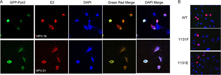
FIG 5.
Nuclear localization of HPV E2 and Pyk2. (A) CV-1 cells were transfected with GFP-Pyk2 WT and FLAG-HPV-31 or -16 E2 constructs. Immunofluorescence staining for HPV-E2 proteins with M2 (FLAG) antibodies and DAPI (blue) was performed. Cells were visualized under ×60 magnification using confocal microscopy. Pyk2 (Green), E2 (red), and DAPI (blue) are indicated. (B) HeLa cells were transfected with FLAG-HPV-31 E2 WT or Y131 mutants. Immunofluorescence staining was done with M2 (FLAG, red) antibodies or DAPI (blue). Magnification, ×60.

