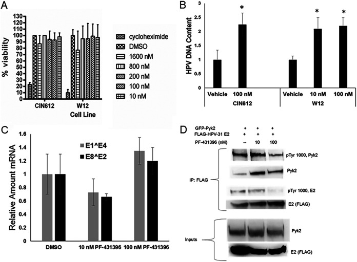
FIG 7.
Pyk2 inhibition increases HPV DNA content by decreasing E2 tyrosine phosphorylation. (A) CIN612-9E and W12 cells were treated with the Pyk2 inhibitor PF-431396 for 24 h, and cell viability was quantified by an MTS cell proliferation assay. Values are expressed as means ± the SEM (n = 8). *, P < 0.05. (B) CIN612-9E cells were incubated with 100 nM PF-431396, and W12 cells were incubated with 10 or 100 nM PF-431396 for 72 h. DNA was isolated, and real-time-PCR was performed for HPV-31 or HPV-16 DNA region near the LCR and normalized to the levels of β-actin. Values are expressed as means ± the SEM (n = 6). *, P < 0.05. (C) CIN612-9E cells were treated with 10 or 100 nM PF-431396 for 72 h. RNA was isolated and converted to cDNA using reverse transcription-PCR. Real-time PCR was carried out with primers to HPV-31 E1̂E4 and HPV-31 E2̂E8 mRNA and normalized to 18S transcripts. Values are means ± the SEM (n = 3). *, P < 0.05. (D) HEK293TT cells were transfected with GFP-Pyk2 and FLAG-HPV-31 E2. Cells were treated with 10 or 100 nM PF-431396 for 24 h. E2 was immunoprecipitated with M2 (FLAG) antibodies. Complexes were blotted with pTyr-1000, Pyk2, and M2 (FLAG) antibodies.

