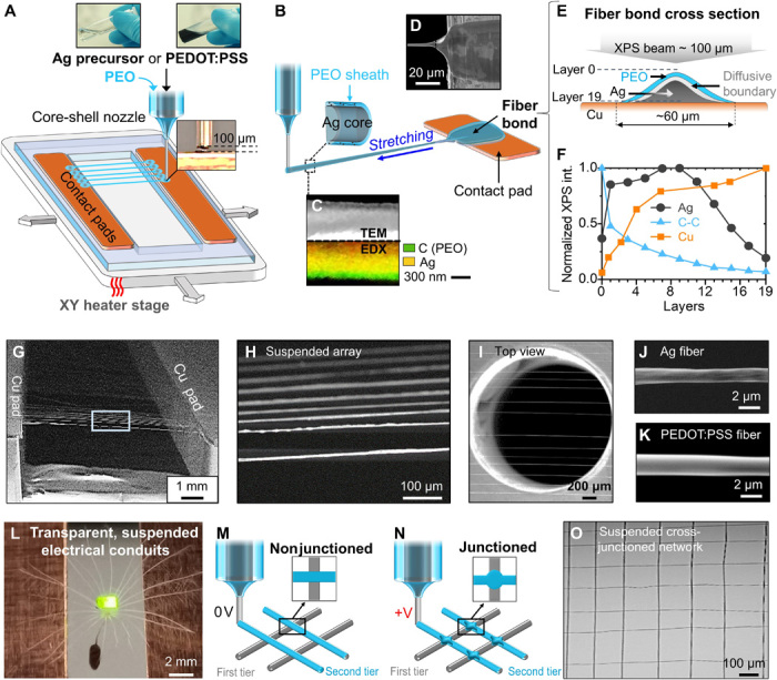
Fig. 1. iFP fabrication of suspended fiber structures with in situ bonding.

(A) Schematic of iFP process for Ag and PEDOT:PSS fibers. (B) Schematic showing a close view of the initiation of the iFP fibers. (C) TEM and EDX of a single Ag fiber. (D) Scanning electron microscopy (SEM) image showing fiber bond from the top view. (E) Cross-sectional schematic of fiber bond. (F) XPS depth profiling on the Ag fiber bond. (G and I) SEM images of typical suspended, aligned fiber array. (J and K) SEM images showing individual Ag and PEDOT:PSS fibers. (L) Image of a powered LED lamp and a dandelion seed on top of a suspended PEDOT:PSS fiber array, with the seed passing through the fiber array (Photo credit: Wenyu Wang, University of Cambridge). (M and N) Schematics of nonjunctioned and junctioned fiber grid structures. (O) Optical image of a suspended iFP fiber network.
