. 2020 Aug 4;19:1102–1119. doi: 10.17179/excli2020-2376

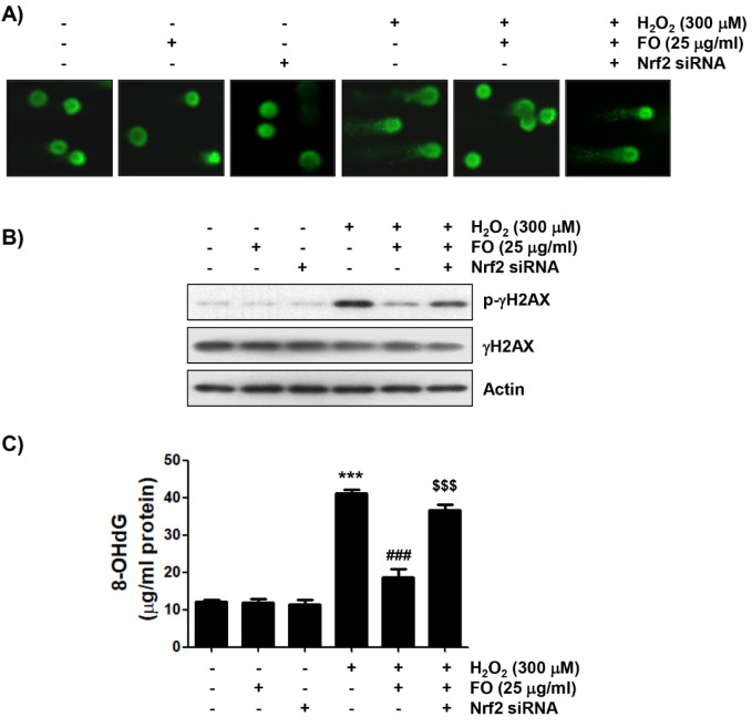
Figure 4. Protection of H2O2-induced DNA damage by FO in MC3T3-E1 cells. Cells transfected with or without Nrf2-siRNA were pretreated with 25 μg/ml FO for 1 h, and then treated with or without 300 μM H2O2 for 24 h. (A) Comet assay was performed, and representative images were captured by fluorescence microscopy (original magnification, ×400). (B) Total cell lysates were prepared, and p-γH2AX and γH2AX expressions were identified by Western blot analysis. The equivalent loading of proteins in each well was confirmed by actin. (C) The DNA samples of cells were subjected to assessment of 8-OHdG level. The measurements were made in triplicate, and the results are expressed by mean ± SD (*** p < 0.001 compared with the control group, ### p < 0.001 compared with the H2O2-treated group, $$$ p < 0.001 compared with the FO and H2O2-treated group).

Figure 4