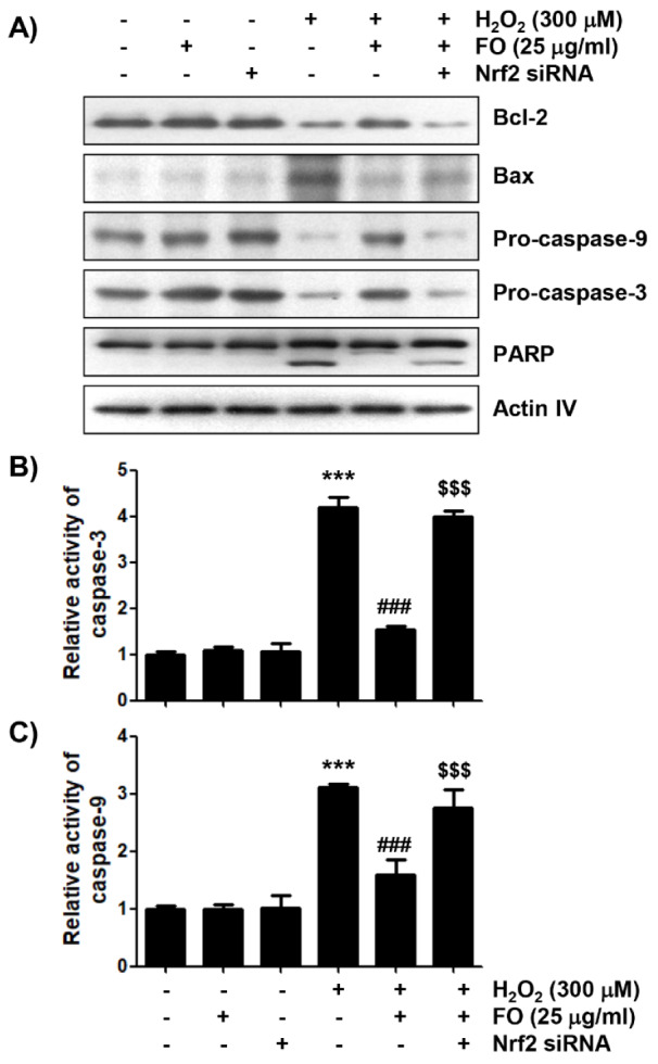
Figure 7. The effects of FO on the expression levels of apoptosis-related proteins, and the activity of caspases in H2O2-treated MC3T3-E1 cells. (A) The cells cultured under the same conditions as Figure 6 were lysed, and Western blot analysis was performed using the indicated antibodies. Actin was used as an internal control. (B) and (C) The activities of caspase-3 and caspase-9 in cell lysates were measured using the respective substrate peptides, Ac-DEVD-pNA and Ac-LEHD-pNA. Data are expressed as the mean ± SD obtained from three independent experiments (*** p < 0.001 compared with the control group, ### p < 0.001 compared with the H2O2-treated group, $$$ p < 0.001 compared with the FO and H2O2-treated group).

