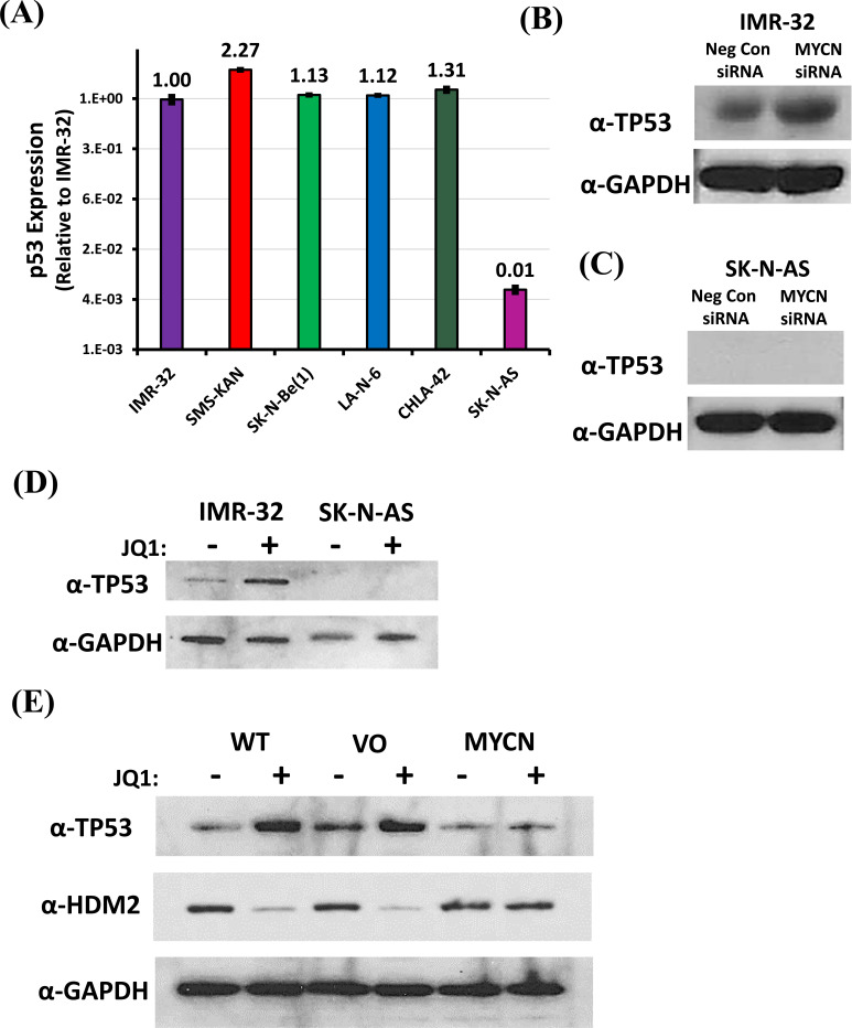
Fig. (5).
Effect of MYCN downregulation on TP53 in neuroblastoma cells. (A) Relative TP53 mRNA expression was acquired from three MYCN-amplified [IMR-32, SMS-KAN, and SK-N-Be(1)] and three non-MYCN amplified (SK-N-AS, LAN-6, and CHLA-42) neuroblastoma cell lines by qRT-PCR. TP53 expression was normalized to GAPDH. Data shown are the composite of triplicate wells from three independent measurements. (B & C) Western blot analysis of the effect on TP53 protein in neuroblastoma cells (IMR-32 and SK-N-AS cells, respectively) treated with MYCN siRNA. Samples include Negative Control siRNA and MYCN siRNA. (D) Western blot analysis of the expression of TP53 protein in neuroblastoma cells (IMR-32 and SK-N-AS cells, respectively) treated with JQ1 (8 µM) compared to untreated cells. (E) Western blot analysis of the expression of TP53 and HDM2 (MDM2) proteins in Wild Type (WT), Vector only control (VO), and exogenously expressing MYCN (MYCN) IMR-32 cells treated with JQ1 (8µM) compared to untreated cells. Total protein analysis was performed compared to GAPDH as a control in all western blots. All data shown are the composite from three independent experiments. (A higher resolution / colour version of this figure is available in the electronic copy of the article).

