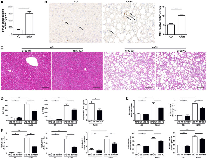
Fig. 2.

MPO deficiency attenuates liver injury in a dietary mouse model of NASH. Male MPO KO and WT littermates were fed with a chow diet (“CD”) or the HFCholC (“NASH”) diet for 24 weeks (nMPO WT CD = 3‐4; nMPO KO CD = 5‐6; nMPO WT NASH = 12‐14; nMPO KO NASH = 15‐17). (A,B) Comparison of chow‐fed and HFCholC‐fed WT mice, respectively. (A) Serum MPO concentrations. (B) Representative hepatic MPO immunohistochemistries (scale bar: 50 μm) and quantification of MPO‐positive cells per view field. (C‐G) Comparison of chow‐fed and HFCholC‐fed MPO KO mice and their WT littermates. (C) Representative hepatic hematoxylin and eosin stainings (scale bar: 100 μm). (D) Serum aspartate aminotransferase and ALT levels and hepatic content of HNE bovine serum albumin. (E) qPCR of apoptosis‐related genes. (F) qPCR of inflammation‐associated genes. Data are presented as mean ± SEM. *P ≤ 0.05, **P ≤ 0.01, ***P ≤ 0.001, ****P ≤ 0.0001. Abbreviations: AST, aspartate aminotransferase; BSA, bovine serum albumin; Bax, Bcl‐2‐associated X protein; Bcl2l11, Bcl‐2‐like protein 11; Bbc3, p53 up‐regulated modulator of apoptosis; Casp8, caspase 8; Ccl2, monocyte chemoattractant protein 1; Il1b, interleukin 1 β; Tnfa; tumor necrosis factor α.
