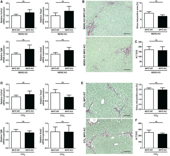
Fig. 5.

MPO is not relevant in nonsteatotic liver fibrosis models. (A‐C) Spontaneous liver fibrosis was evaluated in 62‐week‐old female MDR2‐MPO‐DKO mice and MDR2 KO–MPO WT littermates (n = 11 each). (A) qPCR of fibrosis‐related genes. (B) Representative hepatic sirius red staining (scale bar: 100 µm) and quantification of stained area. (C) Serum ALT. (D‐F) Toxin‐induced liver fibrosis was analyzed in MPO KO mice (n = 8) and WT littermates (n = 9) after 10 intraperitoneal injections of CCl4 over 5 weeks. (D) qPCR of fibrosis‐related genes. (E) Representative hepatic sirius red staining (scale bar: 100 µm) and quantification of stained area. (F) Serum ALT. Data are presented as mean ± SEM. *P ≤ 0.05. Abbreviation: Tgfb, transforming growth factor β.
