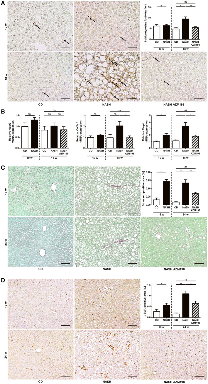
Fig. 7.

Pharmacological MPO inhibition attenuates NASH‐induced liver fibrosis in mice. MPO‐mediated tissue damage and NASH‐induced liver fibrosis were assessed in C57BL/6J mice, fed with chow diet (“CD”) or HFHC diet with (“NASH AZM198”) or without (“NASH”) the MPO inhibitor, AZM198, at different time points: (A,B) Early intervention (nCD 10w = 3‐4; nNASH 10w = 10‐12; nCD 18w = 4; nNASH 18w = 13‐14; nNASH AZM198 = 12‐14). (C,D) Late intervention (nCD 16w = 5; nNASH 16w = 15; nCD 24w = 4; nNASH 24w = 15‐16; nNASH AZM198 = 16). (A) Representative 3‐chlorotyrosine immunostainings of liver sections (scale bar: 50 µm), quantification of stained foci. (B) qPCR of fibrosis‐related genes. Liver fibrosis was quantified histologically by sirius red staining (C) (scale bar: 100 µm) and α‐SMA immunohistochemistry (D) (scale bar: 100 µm). Data are presented as mean ± SEM. *P ≤ 0.05, **P ≤ 0.01, ***P ≤ 0.001.
