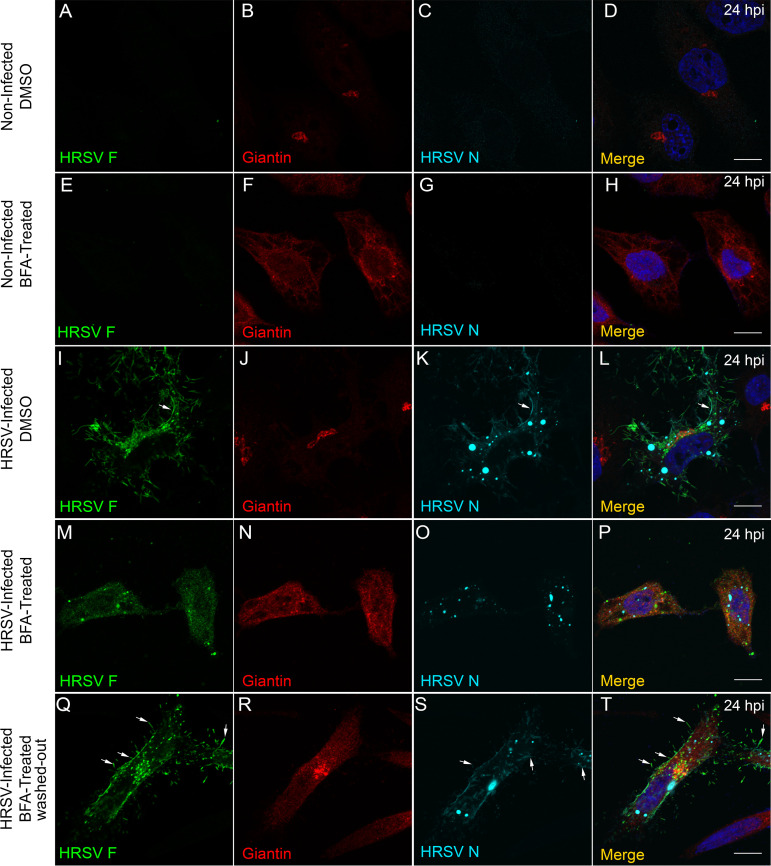
FIG 4.
Treatment with brefeldin A affects the distribution of HRSV F and N proteins. (A) Negative control showing lack of staining for HRSV F in noninfected cells. (B) Intact Golgi upon treatment with DMSO used as vehicle for BFA. (C) Negative control showing staining for HRSV N in noninfected cells. (D) Merge of panels A, B, and C. (F) Effect of BFA on the Golgi morphology, in noninfected cells in panels E, G, and H. (I to L) Localization of HRSV F and N proteins at 24 hpi without BFA treatment. (M to P) Dramatic changes in the localization of HRSV F (M) and N (O) proteins upon BFA treatment. (Q to T) The cell was treated with BFA for 5 hours, and then fresh medium was replaced without BFA; the sizes of the inclusion bodies (S) went back to being similar to the control (K), and it is possible to see filament formation budding from the plasma membrane, indicated by the arrows in panels Q, S, and T. The images represent a single focal plane in two independent experiments. Images were taken with Zeiss 780 confocal microscope. Magnification, ×63. All the scale bars = 10 μm.

