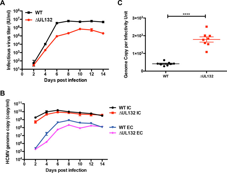
FIG 1.
Decreased virus production from ΔUL132 HCMV-infected cells. (A) Infectious virus production from the extracellular supernatant. Confluent HFFs on 35-mm dishes were infected with either WT or ΔUL132 HCMV at an MOI of 0.1. At the indicated days postinfection (dpi), extracellular infectious virus was quantified as described in Materials and Methods. (B) Intracellular and extracellular HCMV genome copy numbers. Genome copy numbers were determined in the supernatants (extracellular [EC]) and from infected cells (intracellular [IC]) by qPCR of monolayers as described in Materials and Methods. Data are presented as the means and standard errors of the means (SEM) from three independent experiments. (C) HCMV particle-to-infectivity ratios. WT and ΔUL132 HCMV particle-to-infectivity ratios were calculated from extracellular virus at 7 dpi (n = 8); error bars denote SEM. Significances of differences between groups (n = 8 per group) were determined using the Student t test (****, P < 0.0001).

