FIG 6.
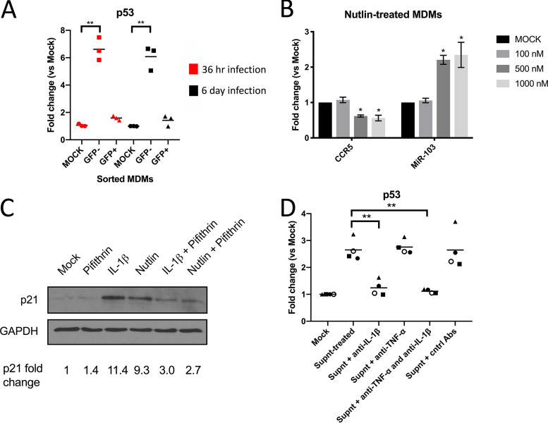
IL-1β-triggered p53 activity modulates CCR5 mRNA and miR-103 levels. (A) Expression of p53 mRNA was determined in sorted macrophages derived from 3 blood donors by real-time qPCR. Bars shown are the mean fold changes compared to mock cells (n = 3 blood donors; Wilcoxon matched-pairs signed-rank test). (B) Different concentrations of the MDM2 antagonist Nutlin-3 were added to MDMs (3 different blood donors), and its effect on CCR5 mRNA and miR-103 levels was determined by real-time qPCR. Shown are the mean fold changes ± SD compared to mock cells (n = 3 blood donors; Student’s t test). (C) Control, IL-1β-treated, or Nutlin-treated MDMs were then exposed to Pifithrin (or the vehicle) for 24 h and lysed, and the p53-driven enhanced expression of p21 was determined by Western blotting. Data shown are derived from MDMs of one blood donor (n = 2 blood donors). (D) The conditioned supernatant from infected macrophages was added to new MDM cultures from 4 blood donors. In some cases, the conditioned supernatant was treated with either control goat anti-human IgGs (Cntrl Abs), neutralizing anti-TNF-α, neutralizing anti-IL-1β, or both neutralizing antibodies. The level of p53 mRNA was then measured by real-time qPCR. Bars represent the mean fold changes compared to mock (untreated) cells (n = 4 blood donors; Wilcoxon matched-pairs signed-rank test).

