
Figure 3.
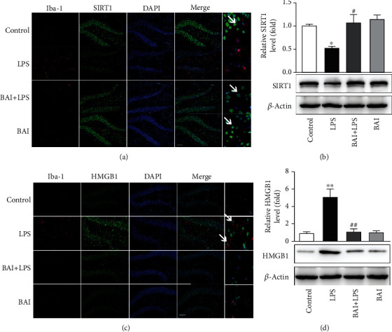
BAI upregulated SIRT1 expression and downregulated HMGB1 expression in mice with hippocampal impairment. (a) Immunofluorescence of SIRT1 (green) in microglial cells (Iba-1+; red) in the mouse hippocampus 24 h after LPS exposure. Arrows point to SIRT1-positive microglial cells (Iba-1+). (b) Western blot analysis of SIRT1 expression in mice subjected to LPS and BAI. (c) Immunofluorescence of HMGB1 (green) in microglial cells (Iba-1+; red) in the hippocampus 24 h after LPS exposure. Arrows point to HMGB1-positive microglial cells (Iba-1+). (d) Western blot analysis of HMGB1 expression in mice subjected to LPS and BAI. Data are expressed as the mean ± SEM (n = 4–5). ∗P < 0.05, ∗∗P < 0.01, control vs. LPS; #P < 0.05, ##P < 0.01, BAI+LPS vs. LPS. SIRT1, silent information regulator 1; HMGB1, high-mobility group box protein 1.