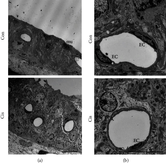
Figure 2.

Cisplatin changes the ultrastructure of SV cells. (a, b) The ultrastructure of SV was observed by TEM. Both the size and the shape of cell nuclei appeared normal, and mitochondria were prevalent and occurred in a highly organized manner in the control mice. A thicker SV and more blood vessels were observed in the cisplatin-treated mice; the numbers of mitochondria and endothelial cells were reduced; and most of the cells were in an abnormal shapes with large and round nuclei, a swollen endoplasmic reticulum, and a thinner vascular wall in cisplatin-treated mice. Con: 0.9% physiological saline; Cis: cisplatin; EC: endothelial cell; m: mitochondrion; PC: pericyte; mu: marginal cell nucleus. Scale bars, 10 μm.
