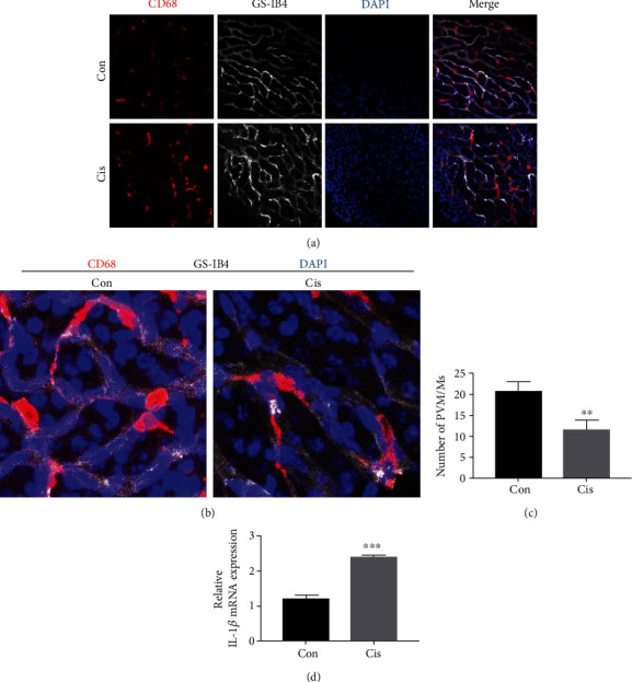
Figure 6.

Cisplatin activates PVM/Ms in SV. (a) The distribution of PVM/Ms by immunofluorescence staining. PVM/Ms were marked with a special antibody against CD68 (red) and strial capillaries with Griffonia Simplicifolia IB4 (gray). Nuclei were stained with DAPI (blue). The PVM/Ms were highly invested on the abluminal surface of capillaries. Scale bars, 100 μm. (b) Zoom in of immunofluorescence staining. (c) Quantification of CD68-positive infiltration per SV area (n = 5). (d) qRT-PCR shows mRNA for IL-1β in the SV. n = 6. ∗∗p < 0.01 and ∗∗∗p < 0.001.
