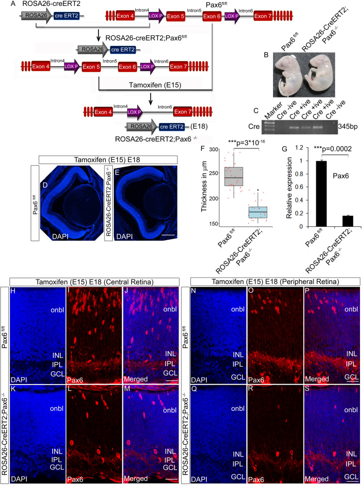
Figure 3.
Generation of Pax6 cKO (Pax6−/−) embryos. (A) Schematic of the breeding strategy used in the study to generate Pax6−/− embryos after tamoxifen injection. (B) Pax6fl/fl and Pax6−/− embryos collected at E18 after tamoxifen injection at E15 display marked phenotypic differences. (C) Genotyping PCR to screen Pax6fl/fl and Pax6−/− animals based on the presence of cre. (D–F) DAPI staining of Pax6fl/fl and Pax6−/− embryos revealed a reduction in the thickness of the neuroretina of Pax6−/− embryos (E) compared to the Pax6fl/fl (D) embryos. Thickness of the retina is plotted as box plot using R program (F; t-test; p < 0.001). Scale bar—25 µm (G) Quantitative expression analysis for Pax6 in Pax6fl/fl and Pax6−/− retina showed a significant reduction in the expression level (p < 0.001). (H–S) Immunostaining with N-terminal Pax6 antibody showed a significant reduction in the number of Pax6 positive cells in both the central (I, L) and peripheral region (O, R) of Pax6−/− retina compared to Pax6fl/fl retina. As such, the numbers of Pax6 positive cells are lesser in the peripheral retina (O) compared to the central retina (I). Scale bar—25 µm. n = 3, at least 3 embryos each of Pax6fl/fl /Pax6−/− from 3 pregnant animals were collected. Error bars indicate SEM from three biological replicates.

