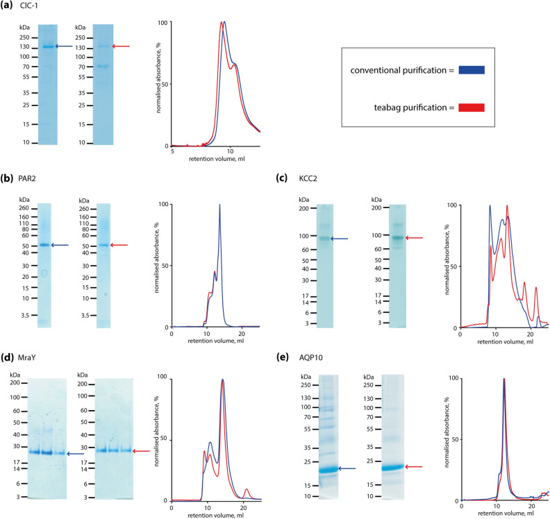
Figure 2.
Comparison of conventional and teabag purification of five different membrane proteins. Each panel shows the results from side-by-side comparison of conventional and teabag purifications, respectively. SDS-PAGE with conventional (left) and teabag (right) purification, and SEC profiles with conventional purification (blue) and teabag purification (red). (a) Human ClC-1 chloride channel; (b) human PAR2; (c) Monodelphis domestica KCC2; (d) Clostridium bolteae MraY; (e) human AQP10. For each pairs of SDS-PAGE images, comparable amounts of protein have been loaded and the brightness and contrast is equal and applied across the entire image, non-cropped SDS-PAGE images are provided in the Supplementary Material.

