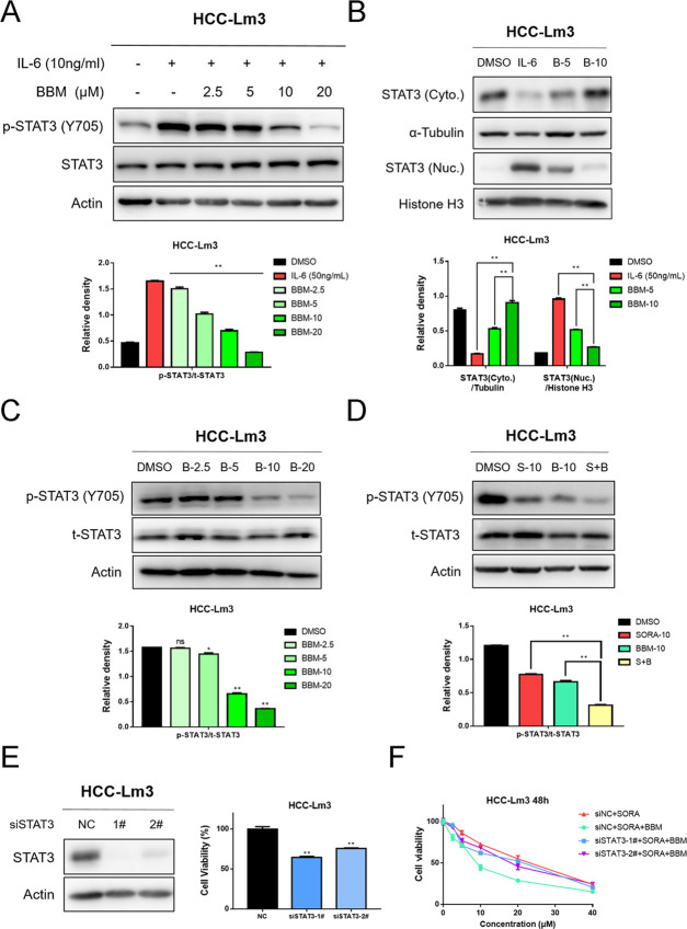
Figure 5.
Combined use of BBM and SORA activities in synergy to suppress STAT3 activation in HCC cells, and knockdown of STAT3 abolished the sensitization effect of BBM. (A) HCC-Lm3 cells were pretreated with 2.5, 5, 10, and 20 μM BBM for 2 h, and then stimulated with IL-6 (10 ng/mL) for 30 min. Western blot analysis was performed to determine STAT3 phosphorylation. Relative STAT3-phosphorylated (Y705) expression (p-STAT3/t-STAT3) was quantified. (B) After HCC-Lm3 cells were treated with IL-6 (10 ng/mL) with or without BBM (5 and 10 μM) for 2 h, western blotting was used to determine the levels of STAT3 in the nucleus and the cytoplasm. (C) HCC-Lm3 cells were treated with BBM at an indicated concentration for 24 h, and then western blot analysis was used to determine the phosphorylated and total STAT3 proteins. (D) Treatment of HCC-Lm3 cells with SORA alone (10 μM), BBM alone (10 μM), or in combination with SORA (10 μM) and BBM (10 μM) for 24 h. Western blotting was used to determine the phosphorylated and total STAT3. The relative expression of all bands in (A)–(D) was quantified using ImageJ software and analysis with Graphpad prism 7. **P < 0.01 compared to the negative control. ns means no significance. (E) STAT3 expression level and cell viability of HCC-Lm3 cells with STAT3 knocking down were examined by western blot analysis and MTS assay. **P < 0.01 compared to the negative control. (F) siNC or siSTAT3 transfected cells were treated with SORA (10 μM) or SORA (10 μM) plus BBM (10 μM) for 48 h, and then the cell viability was measured by MTS assay.

