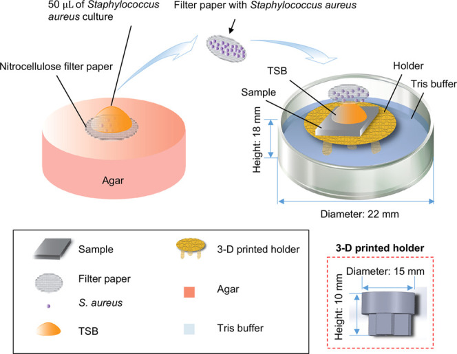
Figure 1.

Schematic illustration of the methods used to study the antimicrobial properties of the Mg-based samples. The red dashed square at the right corner highlights the three-dimensional (3D) printed sample holder and its dimensions. The nitrocellulose filter paper had a diameter to be the same as the width of the samples and fit on top of the square-shaped sample as an inscribed circle to ensure all of the bacteria will be in contact with the sample surface.
