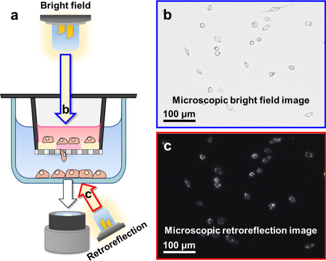
Figure 4.

Evaluation of retroreflection signals from migratory macrophages by comparing different microscopic image fields. (a) Schematic illustration of microscopic image acquisition under bright field (blue arrow, b) and retroreflection (red arrow, c) illumination. (b) Bright-field microscopic images of cells on the surface of the lower chamber. (c) Retroreflection signals from the RJPs inside of cells at the same focus as (b).
