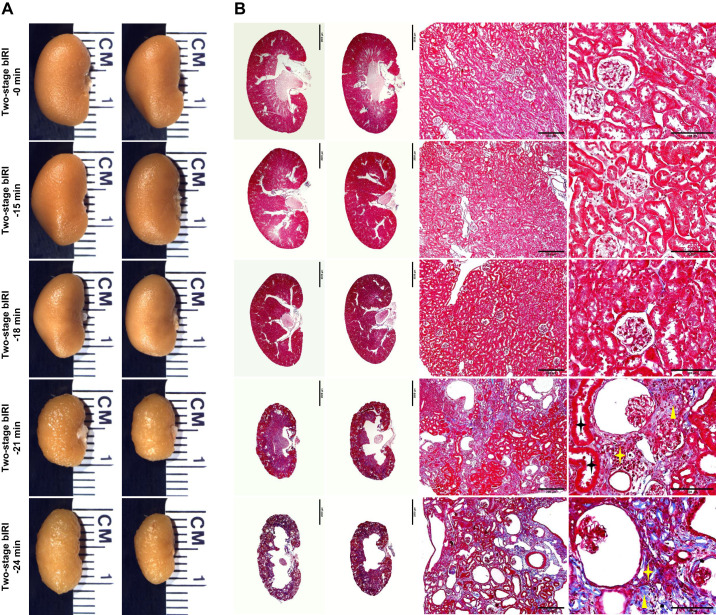
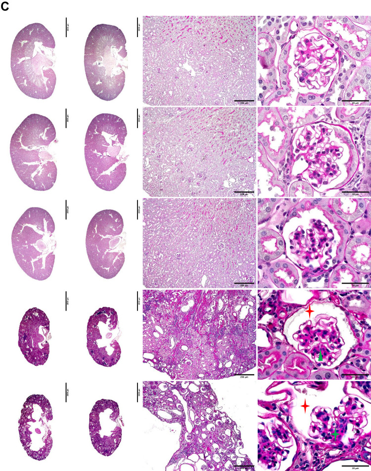
Fig. 2.
Histopathological analyses of the two-stage bilateral ischemia-reperfusion injury (bIRI) model. A: representative images of the left and right kidneys from each two-stage bIRI group. B and C: kidney slices with Masson’s trichrome (B) or periodic acid-Schiff staining (C) showed the typical histopathological features of chronic kidney disease, including tubular atrophy (black stars), interstitial fibrosis (yellow triangles), inflammatory cell infiltration (yellow stars), glomerulosclerosis (green triangle), collapse of glomerular tufts (green star), and dilated Bowman’s capsules (red stars).


