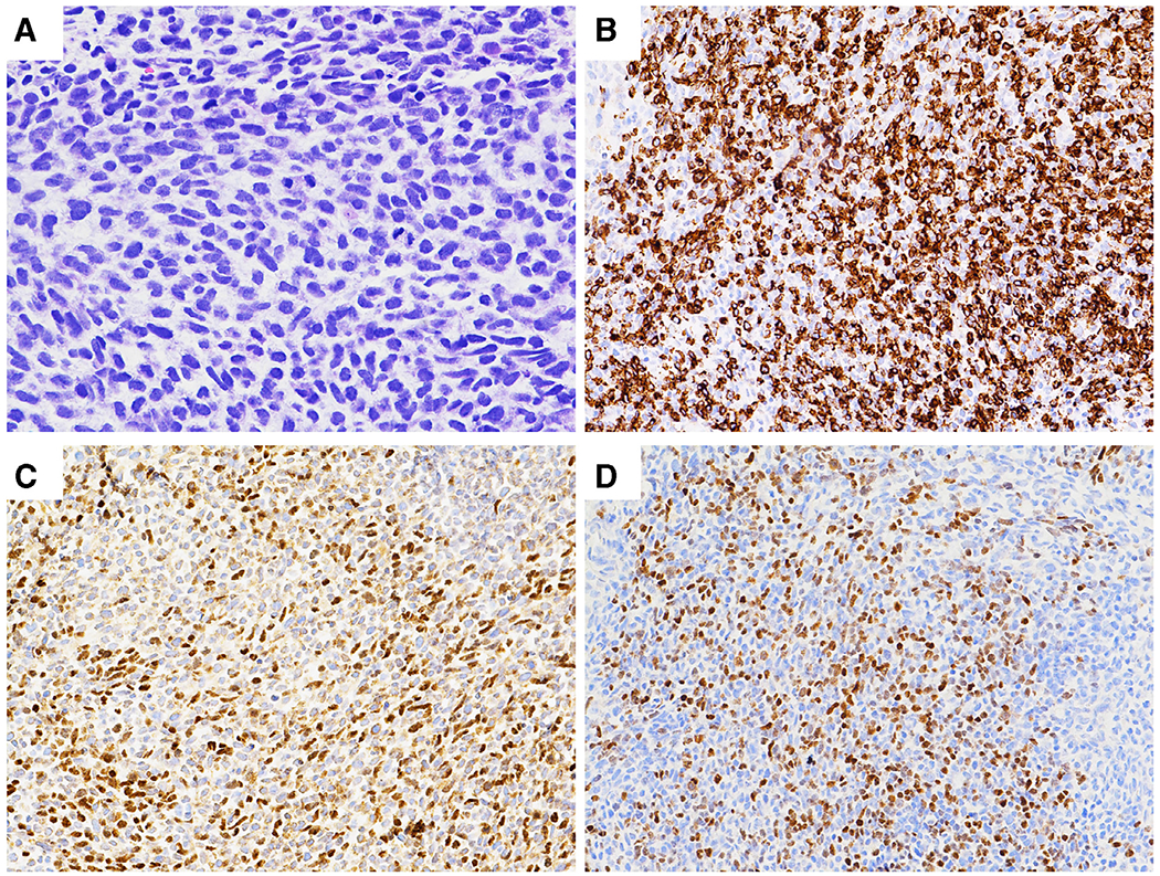
Fig. 1.

Histology and immunostaining results of the patient’s abdominal mass. (A). H&E: A hypercellular tumor with spindle shaped, hyperchromatic and pleomorphic nuclei, coarse chromatin, inconspicuous nucleoli and tapering eosinophilic cytoplasm. Frequent mitoses and karyorrhexis are present. (B). Desmin: Tumor cells were strongly and diffusely positive (cytoplasmic stain). (C). Myo-D: Approximately 35–40% of tumor cells were positive (nuclear stain). (D). Myogenin: Approximately 20% of tumor cells were positive (nuclear stain).
