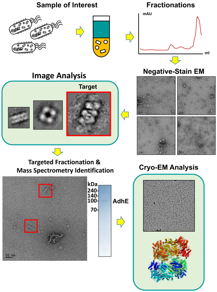
Fig. 1. Schematic workflow of EMPAS: Fractionations, target selection, identification, and structural determination.
Fig. 1. Schematic workflow of EMPAS: Fractionations, target selection, identification, The lysate containing endogenous protein mixtures was fractionated and examined with negative-stain EM. From the collected micrographs, molecular architectures of interest are selected (indicated with red boxes). After target selection, targeted fractionation followed by mass spectrometry revealed the composition of the target molecular architectures. Further cryo-EM analysis determines the high-resolution structure of the targeted molecular architecture (Scale bar = 50 nm, bottom left panel).

