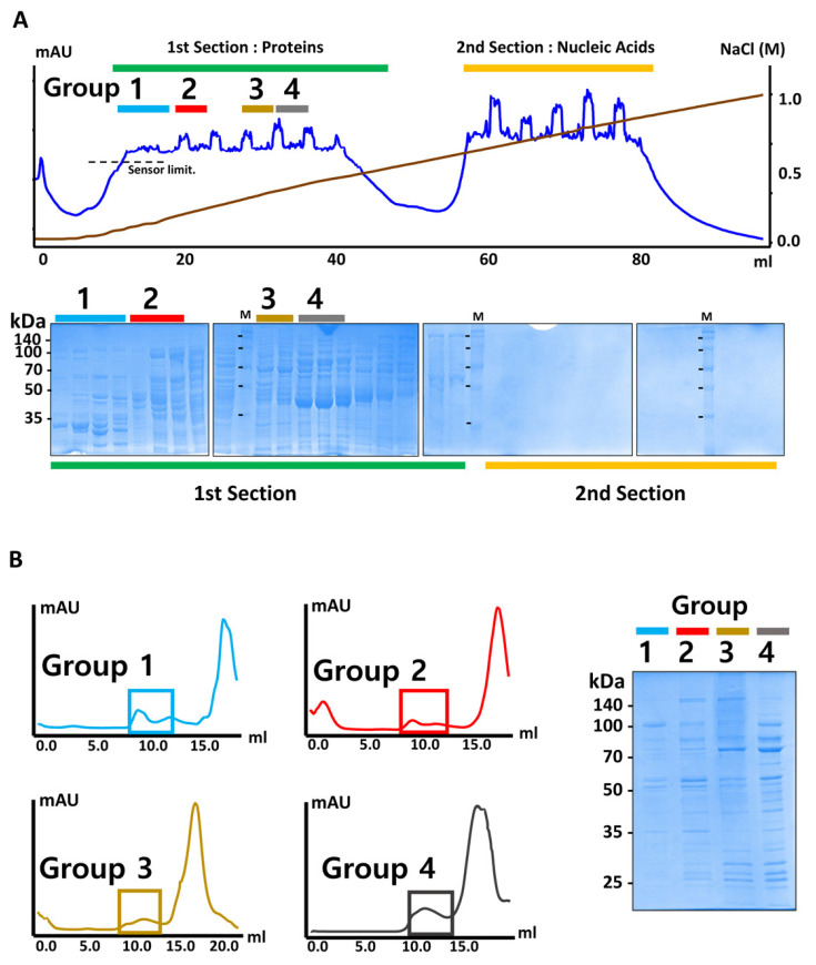
Fig. 2. E. coli sample preparation for EMPAS.
(A) An anion exchange chromatogram profile (top panel) and SDS-PAGE gel (bottom panel) of E. coli cell lysate. The fractionated samples are categorized into four groups based on the pattern of proteins analyzed by SDS-PAGE. (B) Gel filtration profile of each group of the samples (left panel). Proteins eluted at near void volume (boxed) were collected and analyzed by SDS-PAGE (right panel).

