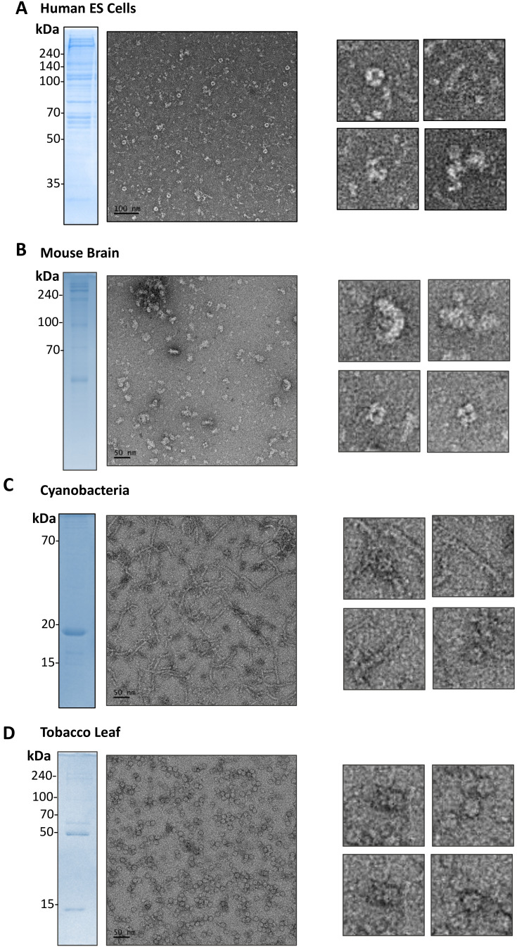
Fig. 5. Representative micrographs of lysates from human stem cells, cyanobacteria, mouse brains and tobacco plant leaves showing the diverse and complex landscape of molecular architectures.
Representative negative-stain EM micrographs (left panels) from fractionated cell lysates of hESCs (A), mouse brains (B), cyanobacteria (C), and tobacco leaves (D) and SDS-PAGE analysis of the sample. A few interesting forms of molecular architectures are shown in the right panels. Scale bars = 50 nm (B-D) or 100 nm (A).

