Figure 2.
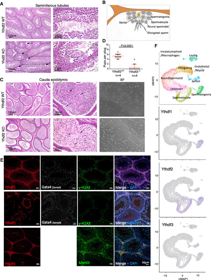
Ythdf2 is essential for male mice fertility. (A) H&E staining showing mild degenerative changes, including scattered vacuoles marked by arrows in the seminiferous tubules in Ythdf2-KO males, compared with WT control. (B) Schematic representation of spermatogenesis inside seminiferous tubules. Differentiation is progressing from spermatogonia at the periphery, via spermatocytes and round spermatids, and ends with elongated mature sperm in the center of the tubule. (C, left) H&E staining of the cauda epididymis, showing severe loss of sperm in Ythdf2-KO compared with control. (Right) Bright field of sperm extracted from the cauda epididymis of KO and control, showing a severe reduction in normal sperm quantity in the KO sample, compared with control. (D) Number of pups per plug produced by mating Ythdf2−/− males, compared with Ythdf2+/− control males. The mothers in both cases were WT. A significant difference between the fertility of KO and heterozygous males is observed (P < 0.0001, Mann–Whitney test). (E) Immunostaining of Ythdf1, Ythdf2, Ythdf3, and Mettl3 in seminiferous tubules, showing that each of the Ythdf proteins is expressed at different stages of spermatogenesis. Costaining of Gata4 typically marks Sertoli cells, costaining of γ-H2AX marks different cells during early spermatogenesis. (F) Dimension reduction representation of single-cell RNA-seq (UMAP) measured in adult mouse testis, showing mild expression of Ythdf1 and Ythdf3 in spermatogonia and in Sertoli cells, and more substantial expression of Ythdf2 in spermatogonia and in spermatocytes.

