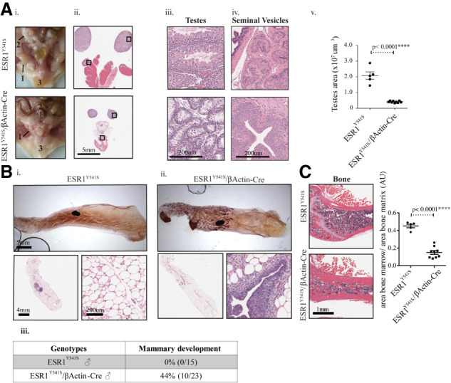
Figure 3.

Male mice expressing ESR1Y541S develop atrophied reproductive organs and display abnormal ductal and bone development. (A, panels i–iv) Pictures and H&E staining of the male reproductive organs at different magnifications showing testes (1), seminal vesicles (2), and preputial glands (3). (Panel v) Quantification of the surface area of the testes in five samples for control mice and nine for experimental mice. (B, panels i,ii) Mammary gland whole mounts of wild-type and experimental male mice as well as H&E sections at different magnifications. (Panel iii) Table summary of the percentage and number of male mice that develop a ductal tree in at least one mammary gland. (C) H&E staining of bone tissue and quantification of the area of bone marrow/area of bone matrix in five control samples and nine experimental samples.
