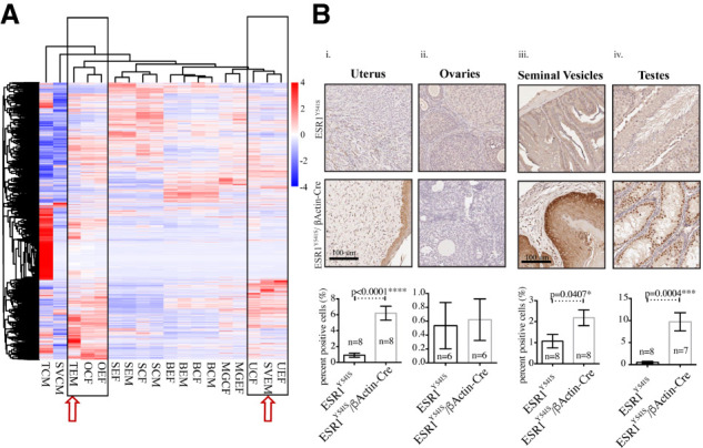
Figure 4.

The reproductive organs of experimental male mice are more transcriptionally similar to female reproductive organs. (A) Heat map of differentially expressed genes. (T) testes, (SV) seminal vesicles, (O) ovaries, (S) spleen, (B) bone, (MG) mammary gland, (U) uterus/fallopian tubes, (E) experimental, (C) control, (F) female, (M) male. (B, panels i,ii) Immunohistochemistry for Sox9 in the uterus/fallopian tubes and the ovaries. (Panels iii,iv) Immunohistochemistry for Sox9 in the seminal vesicles and the testes. Analysis was done using Halo; moderate and strong staining levels were used to quantify the uterus, ovaries, and testes, and strong staining levels were used to quantify the seminal vesicles.
