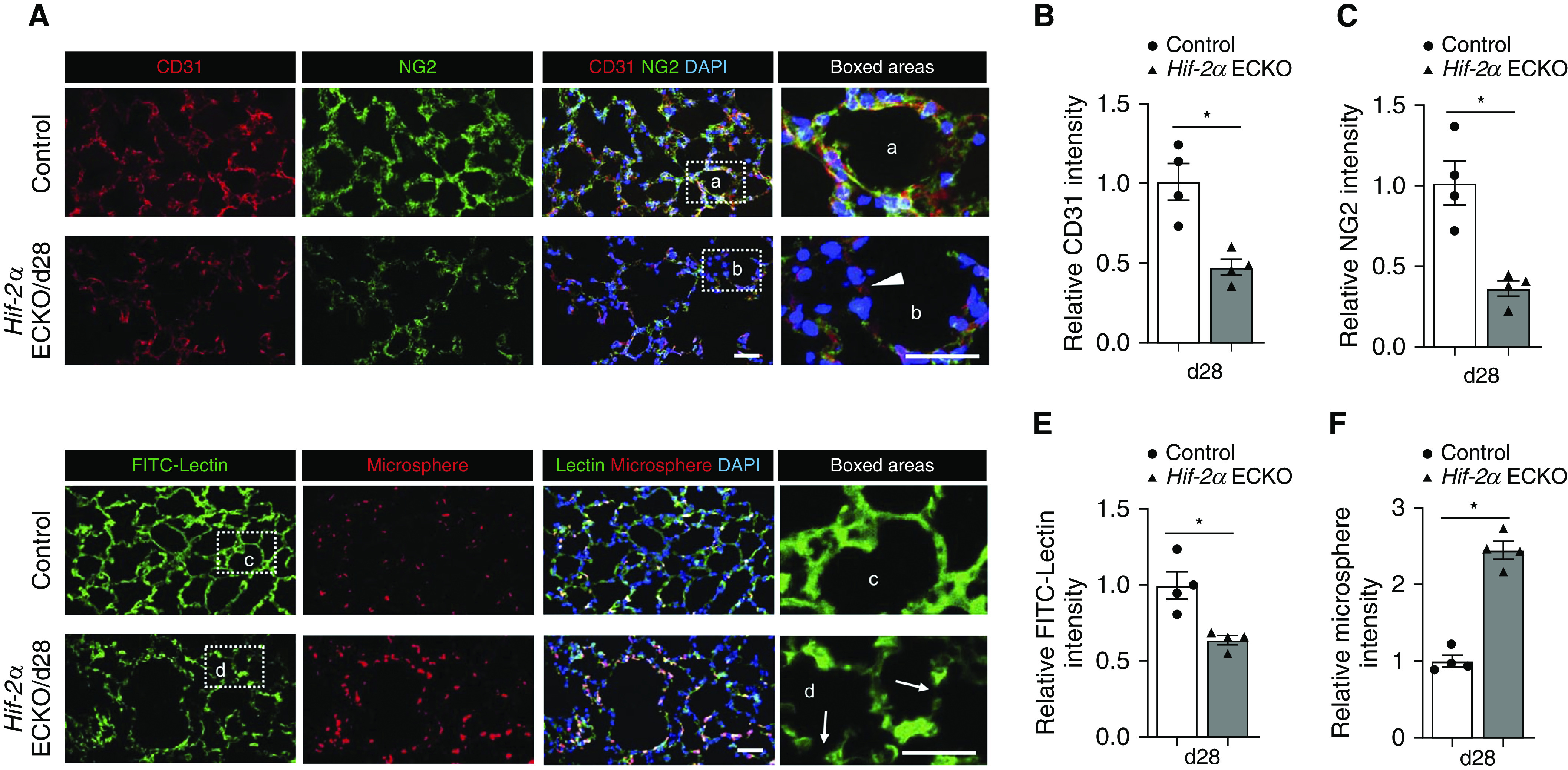
Figure 2.

Induced loss of endothelial Hif-2α (hypoxia-inducible factor-2α) damages alveolar microvasculature. (A) Representative immunofluorescence staining of lung sections from control and Day 28 (d28) endothelial-cell HIF-2α–knockout (Hif-2α ECKO) mice. CD31 (red) identifies endothelial cells, and NG2 (green) identifies pericytes. The white arrowhead in the inset points to an area with low CD31 and NG2 staining in the alveoli of Hif-2α ECKO mice. DAPI (blue) stains the nucleus. (B and C) Quantification of CD31 (B) and NG2 (C) intensity comparing groups shown in A (n = 4). (D) Representative pulmonary microvascular fluorescein isothiocyanate (FITC)-lectin perfusion (green) and microsphere permeability (red) images in control and d28 Hif-2α ECKO mice. White arrows in the inset identify disrupted vasculature in the alveoli of Hif-2α ECKO mice. DAPI (blue) stains the nucleus. (E and F) Quantification of FITC intensity (E) and microspheres (F) comparing groups shown in D (n = 4). Data are presented as mean ± SEM. *P < 0.05 by Mann-Whitney test. Scale bars, 40 μm.
