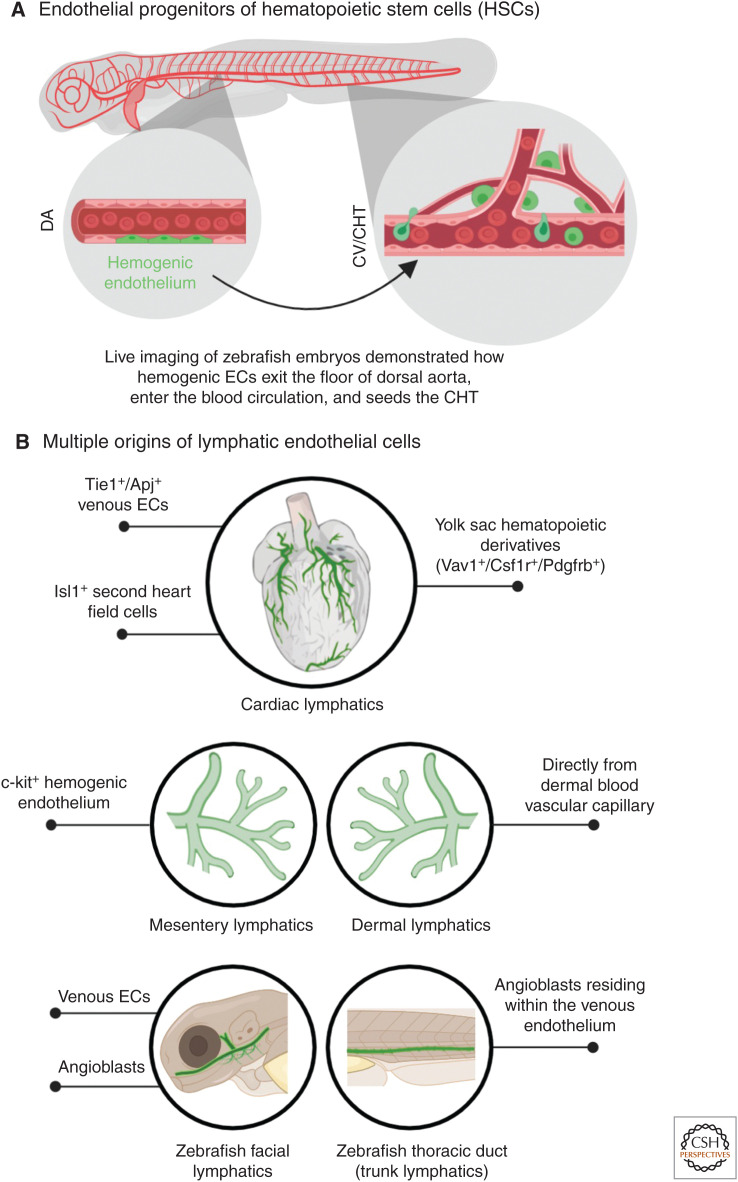
Figure 3.
Origins of hematopoietic stem cells (HSCs) and lymphatic endothelial cells (ECs). (A) Live imaging of zebrafish embryos revealed the existence of hemogenic ECs (green) in the dorsal aorta (DA), which migrate to the caudal end of the embryo and seeds in the cardinal vein (CV), also called caudal hematopoietic tissue (CHT). Here, these cells function as the HSCs. (B) Different progenitor populations that generate organ-specific lymphatic vessels (green) have been revealed through lineage-tracing experiments over the years.

