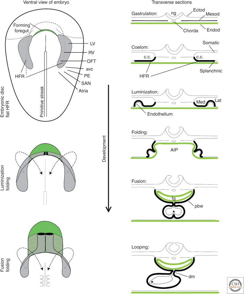
Figure 2.
Early heart development and morphological changes. On the left, it is illustrated that the folding of the embryo associates with foregut formation (green) and that the classically recognized heart-forming region ([HFR], gray, including first heart field [FHF] and second heart field [SHF]) swings toward ventral and medial to progressively fuse at the midline. Within the HFR are cells of the future left ventricle (LV), right ventricle (RV), and the outflow tract (OFT). Contiguous to this HFR are cells that will come to form the atrioventricular canal (avc), proepicardium (PE), the atria, and the sinus venosus/sinus node (SAN). On the right, the illustrations show schematic transverse sections of the changes that occur in association with the folding of the embryo. Note the relation between the lateral HFR rim and the ventral heart tube. The medial HFR becomes the pericardial back wall (pbw), or SHF. (AIP) Anterior intestinal portal, (c.c.) coelomic cavity, (dm) dorsal mesocardium, (Ectod) ectoderm, (Endod) endoderm, (fg) foregut, (Lat) lateral, (Med) medial, (Mesod) mesoderm, (ng) neural groove. The asterisk indicates contact between endocardium and myocardium. (Modified from van den Berg and Moorman 2009, with permission from the authors. Fate-mapping of structures based on data in Bressan et al. 2013 and Dyer and Kirby 2009.)

