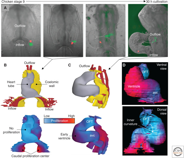
Figure 4.
Additions of cells to the heart tube by cells of the coelomic wall. (A) Fluorescent labeling of cells in a cultured chicken embryo (Hamburger–Hamilton stage 9) to track the substantial movement of cells from the highly proliferative coelomic wall. (B) In Hamburger–Hamilton stage 10, corresponding to the second image from the left in A, the linear heart tube shows virtually no proliferation (1 hour after BrdU exposure), whereas the coelomic wall is highly proliferative. (C) At Hamburger–Hamilton stage 14, corresponding approximately to rightmost image in A, proliferation is starting to reinitiate on the outer curvature of the looped heart tube (1 hour after BrdU exposure). (D) At Hamburger–Hamilton stage 12, long exposure to BrdU (10 hours) reveals pronounced differences in proliferation between the forming ventricle (high, ventral view) and the inner curvature (low, dorsal view). (avc) Atrioventricular canal, (OFT) outflow tract. (A–C based on modified images and 3D models in van den Berg et al. 2009; D based on a 3D model in Soufan et al. 2006.)

