Figure 2.
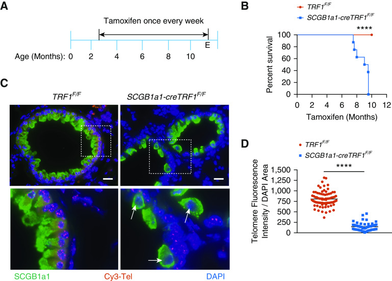
The deletion of TRF1 (telomere repeat binding factor 1) in club cells leads to short telomeres and increased mortality. (A) Tamoxifen-inducible cre-mediated recombination approach to delete TRF1 in cre-expressed cells. Tamoxifen was administered intraperitoneally to mice weekly starting from 10 weeks of age at 250 mg/kg body weight. (B) Kaplan-Meier survival graph of TRF1F/F (telomere repeat binding factor 1 flox/flox) and SCGB1a1-creTRF1F/F mice treated with weekly injections of tamoxifen at 250 mg/kg body weight. ****P < 0.0001 (log-rank test). (C) Telomere quantitative fluorescence in situ hybridization assay conducted on paraffin-embedded lung sections collected after 8–9 months of tamoxifen administration. Cy3-Tel is a PNA probe to detect TTAGGG repeats. SCGB1a1 was used as an antibody to detect club cells. DAPI was used to detect nuclei. The boxed area of the upper panels is shown enlarged in the bottom panels. White arrows point to cells depicted in the boxed area. Scale bars, 20 μm. (D) Quantification of telomere length using telomere fluorescence intensity with reference to DAPI area. N = 3 mice/group. Each dot represents telomere fluorescence intensity in each club cell acquired from twenty different high-power fields. Data are pooled from three mice. ****P < 0.0001 (t test). E = endpoint; PNA = peptide nucleic acid.

