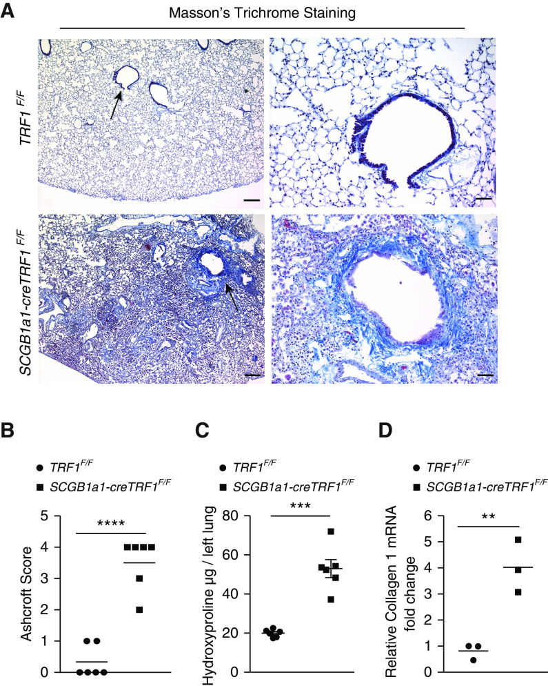
Figure 3.
Airway-centric lung remodeling and fibrosis. (A) Masson’s trichrome staining was performed on lung sections from TRF1F/F and SCGB1a1-creTRF1F/F mouse lungs treated with weekly doses of tamoxifen at 250 mg/kg body weight. Deposition of collagen is indicated by blue stain. Arrows indicate areas of collagen deposition around bronchioles. Scale bars, 400 μm (left panels) and 100 μm (right panels). (B) Quantification of fibrosis using Ashcroft score. ****P < 0.0001 (t test). (C) Quantification of collagen deposition by hydroxyproline assay on lung tissues from TRF1F/F and SCGB1a1-creTRF1F/F mice. ***P < 0.001 (t test). (D) Quantitative PCR of collagen1 mRNA in lungs from TRF1F/F and SCGB1a1-creTRF1F/F mice 9 months after tamoxifen administration. **P < 0.01 (t test).

