
Figure 5.
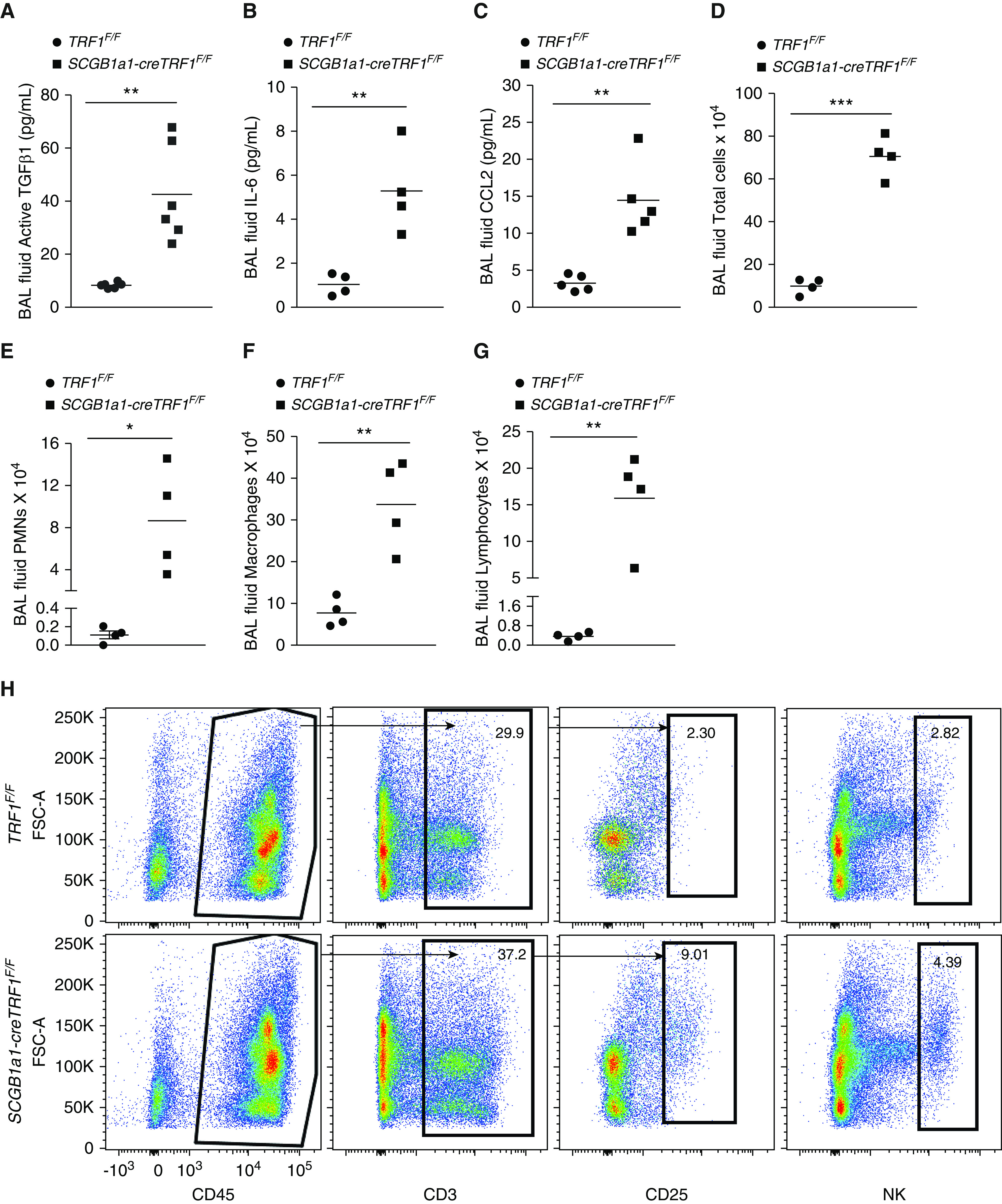
Mouse BAL fluid ELISA, cell count differential analysis, and lung immune cell analysis. (A–C) ELISA on BAL samples from TRF1F/F and SCGB1a1-creTRF1F/F mice to test for active TGF-B1 (A), IL-6 (B), and CCL2 (C). (D–G) Cell count and differentials of BAL fluid samples from lungs of TRF1F/F and SCGB1a1-creTRF1F/F mouse lungs treated with weekly doses of tamoxifen for 9 months; total cell count (D), PMNs (E), macrophages (F), and lymphocytes (G). N = 4 mice/group. *P < 0.05, **P < 0.01, and ***P < 0.001 (t test). (H) Immunophenotyping of lungs from TRF1F/F and SCGB1a1-creTRF1F/F at 9 months by flow cytometry. Percentage of CD3, CD25, and NK cell populations were derived from CD45-positive gate. Black arrows indicate the gating directionality. N = 3. Individual population numbers are mentioned in Table E1. FSC = forward scatter; NK cell = natural killer cell; PMNs = polymorphonuclear cells.