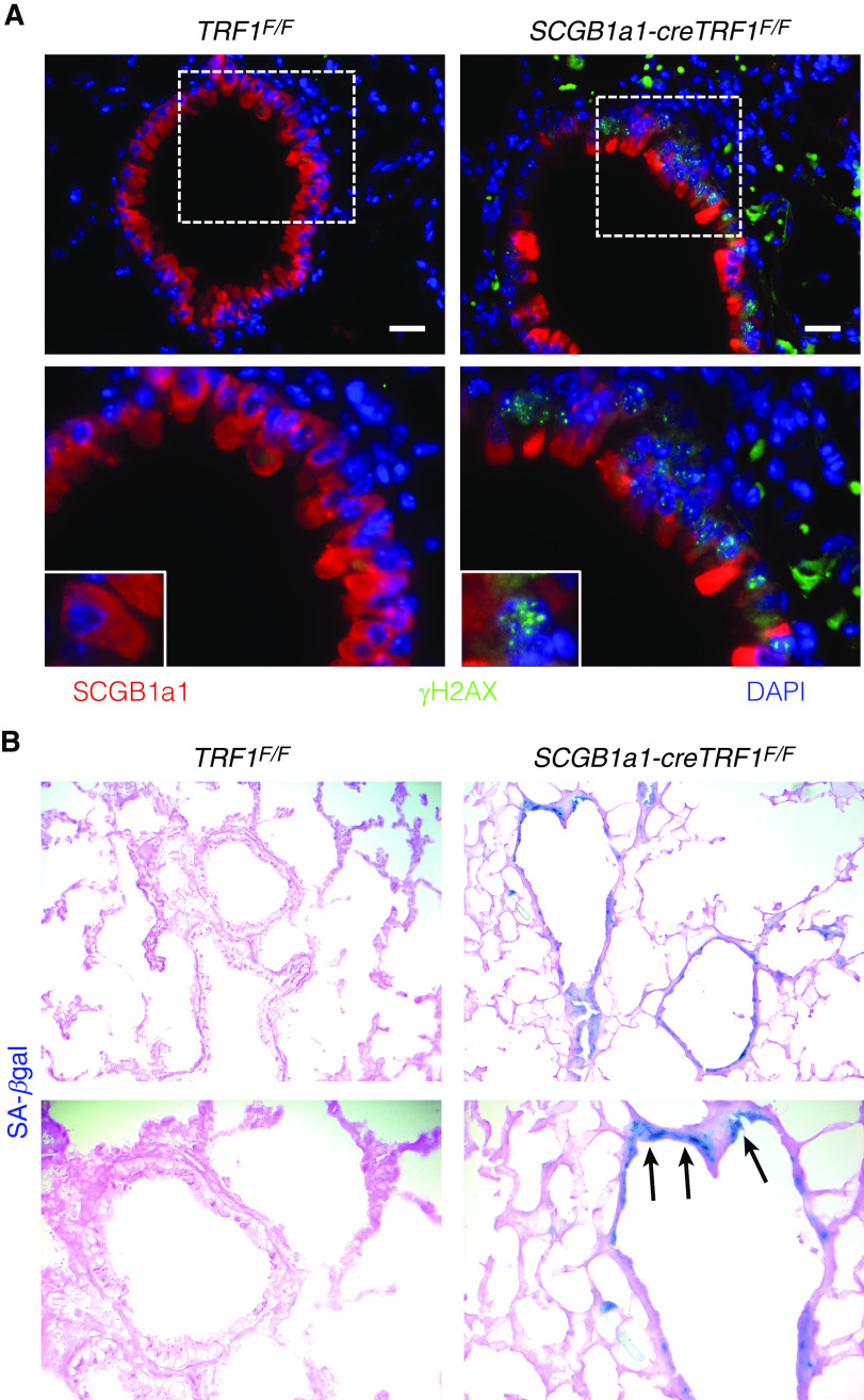
Figure 6.
Immunostaining of lung sections and detection of cellular senescence by SA-β-gal activity. (A) Immunostaining on sections of lungs harvested from TRF1F/F and SCGB1a1-creTRF1F/F mice treated with weekly tamoxifen injections for 9 months. SCGB1a1 immunostaining for club cells (red) and γH2AX (green). γH2AX foci (green) in the nuclei of club cells. Nuclei were stained with DAPI (blue). Scale bars, 20 μm. Boxed area is enlarged in bottom panel along with inset showing individual club cell. (B) SA-β-gal staining of sections of lungs harvested from TRF1F/F and SCGB1a1-creTRF1F/F mice after treatment for 9 months with weekly injections of tamoxifen (250 mg/kg body weight). Note the blue SA-β-gal+ cells (arrows) in lungs of SCGB1a1-creTRF1F/F mice.

