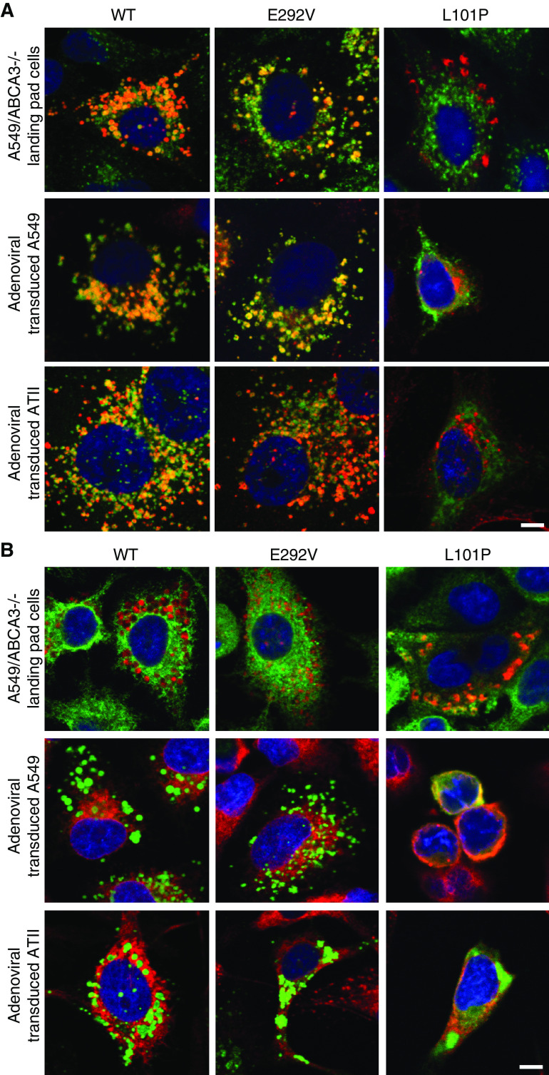
Figure 2.
ABCA3 immunofluorescence colocalization results for A549/ABCA3−/− cells that express ABCA3 WT, p.E292V, or p.L101P are similar to results from adenoviral transduced A549 and ATII cells. Single x-y–plane confocal immunofluorescence colocalization demonstrates that ABCA3 WT and type II (impaired phospholipid transport) mutant p.E292V colocalize with (A) the lysosomal marker (anti-CD63), whereas type I (mistrafficking) mutant p.L101P colocalizes with (B) the endoplasmic-reticulum (ER) marker SelectFX_ER. Of note, in A549/ABCA3−/− cells, ABCA3 (or mutant) is labeled with mCherry (red), anti-CD63 (green), and SelectFX_ER (green). In adenoviral transduced A549 and ATII cells, ABCA3 (or mutant) is labeled with GFP (green), anti-CD63 (red), and SelectFX_ER (red). Scale bars, 10 μm. These experiments were performed three times with similar results.

