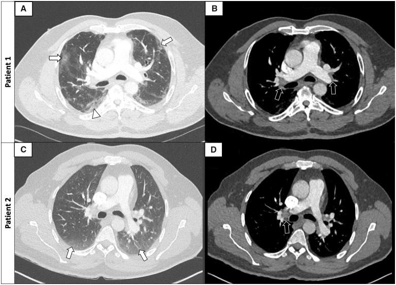
Figure 2.
Examples of axial CTPA images of PE in COVID-19 patients (lung windows: A–C; mediastinum windows: B–D). Patient 1: 42-year-old male with peripheral ground-glass opacities of 25–50% (A, arrows) with small areas of consolidation (arrowhead) and bilateral proximal PE (B, arrows). Patient 2: 57-year-old male with peripheral ground-glass opacities <25% (C, arrows) and right proximal PE (D, arrow). CTPA, computed tomography pulmonary angiography; PE, pulmonary embolism.

