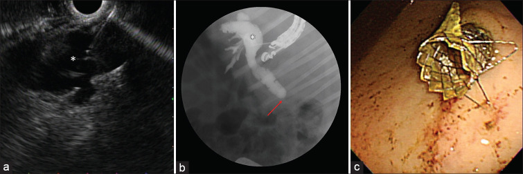
Figure 4.
EUS-guided hepaticogastrostomy: (a) EUS showing dilated left hepatic duct (*) accessed with a 19 G needle, (b) fluoroscopic view showing the 19 G needle in the dilated left hepatic duct (*) and subsequent cholangiography showing dilated common bile duct with distal biliary stricture (arrow), (c) endoscopic view from the stomach showing the distal end of a fully covered self-expandable metal stent

