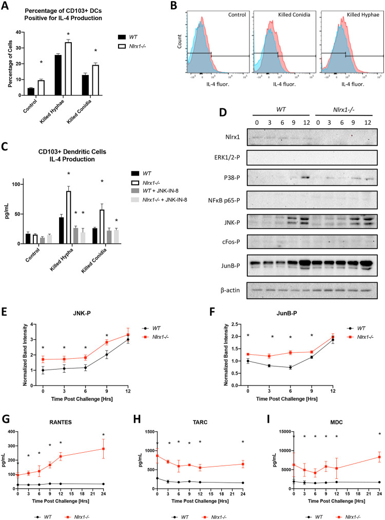
Fig 6. Temporal immune responses by CD103+ DCs during in vitro fungal challenge assays.
Freshly harvested AF293 conidia (5 X 10⁵) were challenged against wild type and Nlrx1-/- CD103+ DCs (5 X 10⁵) at 37°C at 5% CO2. Supernatant and cells were harvested immediately prior to challenge (0 hrs), and at 3, 6, 9, 12 hrs post challenge. (A) Percentage of wild type and Nlrx1-/- CD103+ DCs staining positive for IL-4 12 hours post treatment with killed hyphae and conidia. (B) Representative histograms and gating for IL-4 fluorescence. See S2 Fig for strategy. (C) Intracellular measurement of IL-4 by wild type and Nlrx1-/- CD103+ dendritic cells 12 hours post treatment with killed hyphae and conidia in the presence and absence of the JNK inhibitor (JNK-IN-8). (D) Western blot analysis of phosphorylated (-P) ERK1/2-P, P38-P, NFκB p65-P, JNK-P, cFos-P, JunB-P, Nlrx1, and β-actin from wild type and Nlrx1-/- CD103+ dendritic cells during a 12-hour challenge against viable A. fumigatus conidia. Quantification of (E) JNK-P and (F) JunB-P. Measurement of secreted (G) RANTES (CCL5), (H) TARC (CCL17), (I) MDC (CCL22) by wild type and Nlrx1-/- CD103+ dendritic cells during a 24-hour challenge against viable A. fumigatus conidia. Asterisk denotes statistical significance, P < 0.05 Mann-Whitney U test. Error bars indicate standard deviation. N = 6–8 per experimental group.

