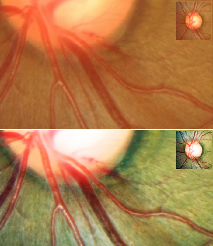
Fig 4. Representative optic-disc photography (ODP) of eye with optic disc hemorrhage (DH).
(A) Magnified image of inferotemporal area in original high-resolution ODP, (B) Magnified image of inferotemporal area in deep-learning-based enhanced ODP. The enhanced ODP improved the color and spatial contrast between the DH and the background retinal color.

