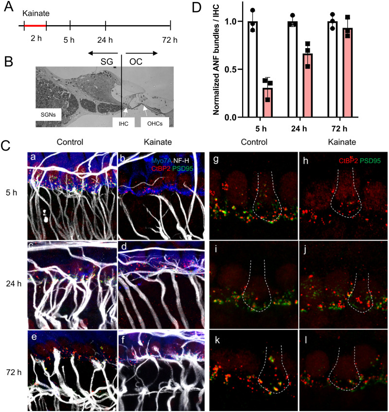
Fig 1. Synaptic regeneration in cochlear explants after kainate treatment.
(A) Timeline of experimental procedure. (B) A cochlear cross section showing the plane of microdissection separating the spiral ganglion (SG) fraction from the Organ of Corti (OC) fraction. (C) The afferent nerve fibers of spiral ganglion neurons, seen with immunostaining for neurofilament (in white), have synapses with hair cells, as revealed by immunostaining for PSD-95 (in green) and CtBP2 (in red) under each hair cell, stained with Myo7A (in blue), after 5 h in culture (Control). Treatment of the mouse cochlea with kainate for 2 hours resulted in massive loss of the peripheral processes and post-synaptic densities of spiral ganglion neurons by 5 h (Kainate). By 24 h, the peripheral processes had grown and formed synapses (characterized by juxtaposition of CtBP2 and PSD-95 immunostaining) with hair cells (Kainate). At 72 h, there was fiber loss in both the control and treated (Kainate) cultures. Panels a-f highlight auditory nerve fibers. Panels g-l highlight synapses; the white dotted line in each panel outlines a hair cell. Scale bars 20 μm. (D) Quantification of auditory nerve fiber (ANF) bundles per inner hair cell after kainate treatment (red bars) normalized to untreated samples (white bars) at the same time point. Data are presented as means ± SD per group. N = 3 explants per group.

