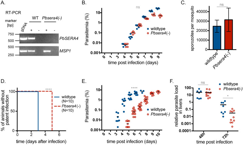
Fig 3. Absence of PbSERA4 delays the time to appearance of Plasmodium-infected red blood cells.
(A) Depletion of PbSERA4 transcripts in Pbsera4(-)-ANKA parasites. cDNA from WT and Pbsera4(-) late-stage schizonts were used to verify the SERA4 specific PCR reaction. MSP1 transcript was used as a positive control. RT-PCR was performed in the presence (+) or absence (-) of reverse transcriptase (RT). Parasite genomic DNA (gDNA) was included as an amplification control. (B) Blood stage growth of Pbsera4(-)-ANKA P. berghei parasites. Naïve NMRI mice (n = 5) were injected intravenously with 1,000 wildtype or 1,000 Pbsera4(-) P. berghei-infected erythrocytes. Infection was then monitored by daily examination of Giemsa-stained blood smears to determine the parasitemia. ns, not significant (unpaired t-test, day 6 values). (C) Development of Pbsera4(-)-ANKA P. berghei in mosquitoes. Sporozoites extracted from salivary glands of infected mosquitoes were quantified 17–25 days after infection. Data is presented as the mean +/- SD. ns, not significant (paired t-test, n = 4 independent experiments). (D and E) Delayed patency following Pbsera4(-)-ANKA sporozoite infection. C57BL/6 mice were infected intravenously with 10,000 sporozoites, and Giemsa-stained blood smears were monitored daily to determine presence of parasites (D) ****, p<0.0001, Log-rank (Mantel-Cox) test, n = 10) and parasitemia (E) ****, p <0.0001 (unpaired t test, day 6 values). (F) Pbsera4(-)-ANKA parasites exhibit no growth defects in infected livers. RNA was extracted from livers of C57BL/6 mice infected with 50,000 to 100,000 sporozoites at the indicated time points. Levels of Pb18s rRNA were normalized to levels of mouse HPRT RNA by qPCR. ns, not significant; *, p < 0.05 (unpaired t-test).

